Patients’ information
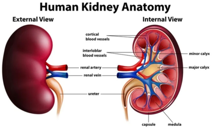
The kidneys are two bean-shaped organs located retroperitoneally on either side of the vertebral column, typically between the T12 and L3 vertebrae. The right kidney is slightly lower than the left due to the position of the liver. Each kidney weighs approximately 120–150 grams in adults.
Structure
Each kidney consists of:
- Renal cortex – the outer region containing glomeruli and convoluted tubules.
- Renal medulla – inner region containing renal pyramids.
- Renal pelvis – funnel-shaped structure that collects urine and channels it into the ureter.
- Nephrons – the functional units (about 1 million per kidney).
Each nephron includes:
- Glomerulus
- Bowman’s capsule
- Proximal convoluted tubule
- Loop of Henle
- Distal convoluted tubule
- Collecting duct
Functions
- Filtration of Blood
The kidneys filter approximately 180 liters of plasma daily. Waste products such as urea, creatinine, and uric acid are removed.
- Regulation of Fluid and Electrolytes
They maintain sodium, potassium, calcium, and phosphate balance.
- Acid-Base Balance
The kidneys regulate blood pH by excreting hydrogen ions and reabsorbing bicarbonate.
- Blood Pressure Control
Through the renin-angiotensin-aldosterone system (RAAS), kidneys regulate blood pressure.
- Hormone Production
- Erythropoietin (stimulates RBC production)
- Renin (blood pressure regulation)
- Activation of Vitamin D (calcium absorption)
Kidney dysfunction can lead to chronic kidney disease, electrolyte imbalance, and systemic complications.
The ureters are narrow, muscular tubes about 25–30 cm long that transport urine from the kidneys to the bladder.
Structure
Each ureter has three layers:
- Inner mucosa
- Middle smooth muscle layer
- Outer adventitia
Function
Urine is propelled downward by peristaltic contractions. There are three natural narrow points where stones commonly lodge:
- Ureteropelvic junction
- Crossing over iliac vessels
- Ureterovesical junction
Obstruction of the ureter can lead to hydronephrosis and severe pain.
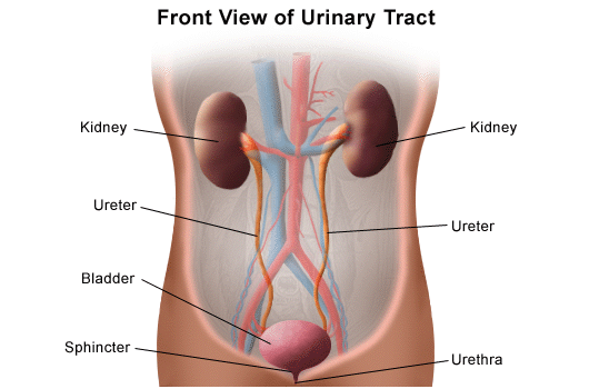
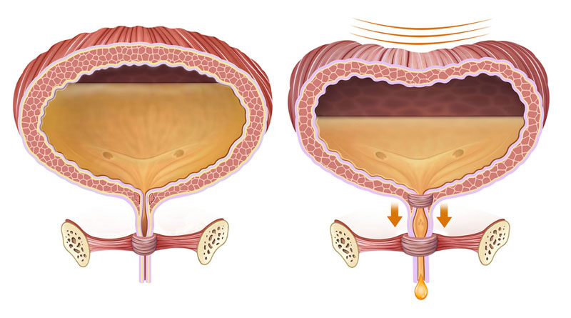
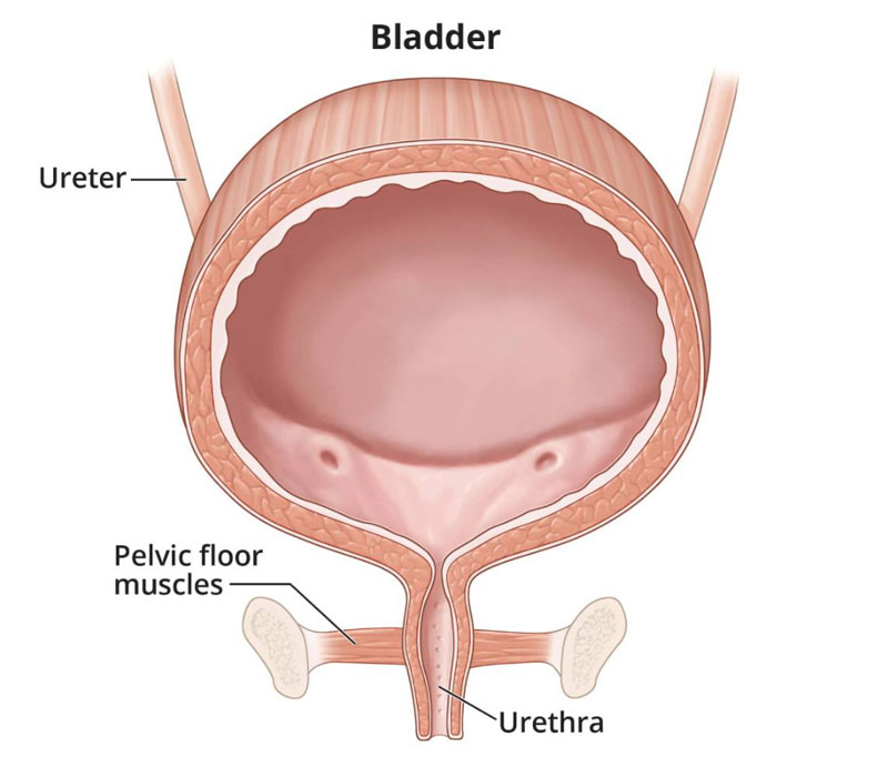
The urinary bladder is a hollow, distensible muscular organ located in the pelvic cavity.
Structure
It consists of:
- Detrusor muscle (smooth muscle layer)
- Trigone (smooth triangular area between ureter openings and urethra)
- Internal urethral sphincter
Function
Storage Phase
The bladder relaxes to store urine (normal capacity 400–600 mL).
Voiding Phase (Micturition)
When full, stretch receptors send signals to the brain. The detrusor muscle contracts and sphincters relax to allow urination.
Bladder control involves both voluntary and involuntary nervous systems.
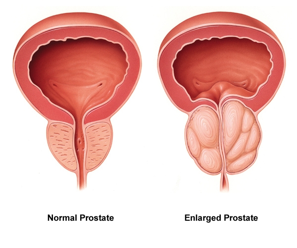
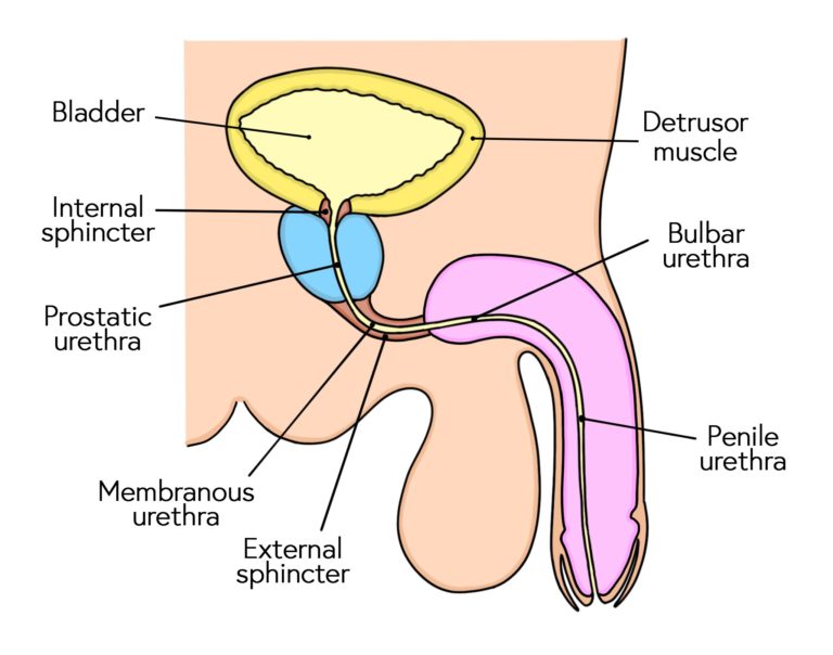
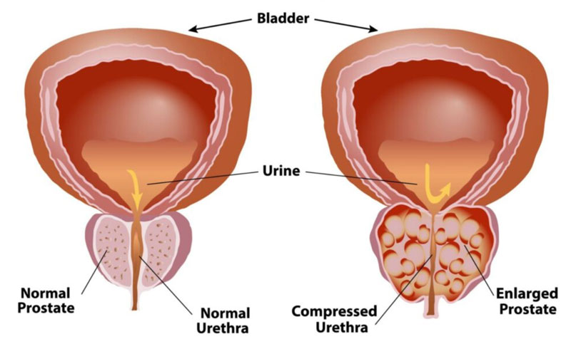
The prostate is a walnut-sized gland found only in males, located below the bladder and surrounding the proximal urethra.
Structure
Divided into:
- Peripheral zone
- Central zone
- Transitional zone
Functions
- Produces alkaline fluid that protects sperm.
- Contributes about 30% of semen volume.
- Helps propel semen during ejaculation.
Common Conditions
- Benign prostatic hyperplasia (BPH)
- Prostatitis
- Prostate cancer
Enlargement can compress the urethra and cause urinary symptoms.
The urethra is a tubular structure that carries urine from the bladder to the exterior.
Male Urethra
Approximately 18–20 cm long.
Divided into:
- Prostatic
- Membranous
- Spongy (penile)
Carries both urine and semen.
Female Urethra
Approximately 4 cm long.
Shorter length makes females more prone to urinary tract infections.
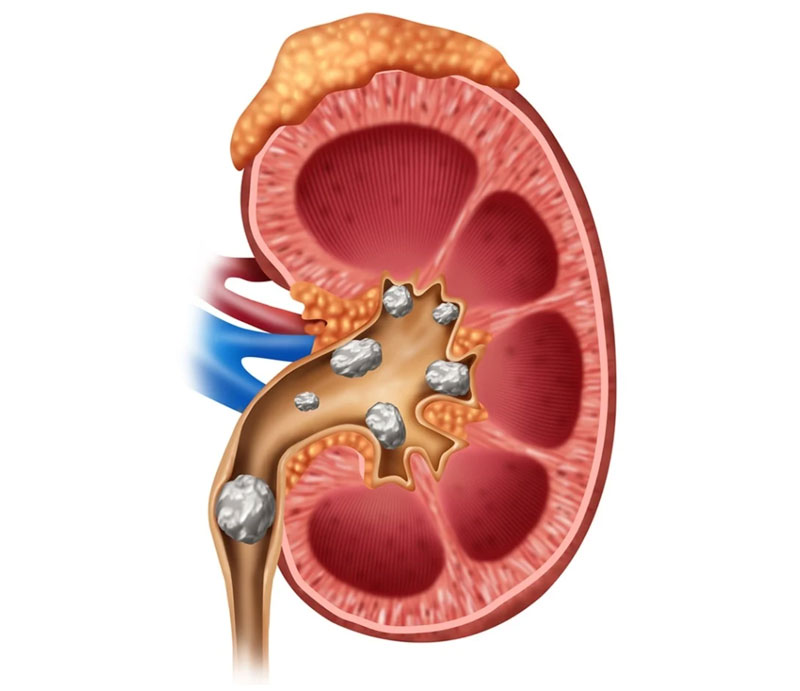
Kidney stone are hard crystalline mineral deposits that form in the kidneys.
Types
- Calcium oxalate (most common)
- Uric acid
- Struvite
- Cystine
Causes
- Dehydration
- High salt diet
- Metabolic disorders
- Family history
Symptoms
- Severe colicky flank pain
- Hematuria (blood in urine)
- Nausea and vomiting
- Pain radiating to groin
Treatment
- Increased fluids
- Pain management
- Lithotripsy
- Surgical removal in severe cases
Prostate Cancer
Prostate cancer is one of the most common cancers in men.
Risk Factors
- Age > 50
- Family history
- Hormonal factors
Symptoms
- Difficulty urinating
- Weak stream
- Bone pain (advanced stage)
Diagnosis
- PSA blood test
- Digital rectal examination
- Biopsy
Treatment
- Surgery
- Radiation therapy
- Hormone therapy
Bladder Cancer
Bladder cancer arises from the bladder lining.
Risk Factors
- Smoking (most significant)
- Chemical exposure
- Chronic irritation
Symptoms
- Painless hematuria
- Frequent urination
- Pelvic pain (late stage)
Diagnosis
- Urine cytology
- Cystoscopy
- Imaging
Treatment
- Tumor resection
- Intravesical therapy
- Chemotherapy
- Radical cystectomy
Urinary dysfunction refers to abnormal urination patterns.
Types
- Urinary Incontinence
Loss of bladder control.
- Stress incontinence
- Urge incontinence
- Overflow incontinence
- Urinary Retention
Inability to empty bladder fully.
- Overactive Bladder
Sudden urge to urinate frequently.
Causes
- Nerve damage (diabetes, spinal injury)
- Enlarged prostate
- Infection
- Aging
- Pelvic floor weakness
Management
- Pelvic floor exercises
- Medications
- Catheterization
- Surgery (in severe cases)